Autism is not a benign difference in brain function. Here, a neuroscientist explains a type of pathology often seen in the brains of autistic individuals — abnormal micro-structural development of the cerebral cortex — and an implication for potential intervention.
Human cortical development between 26 and 39 weeks of gestational age. (Image: Wikipedia)
By Manuel Casanova, MD
Abnormal behaviors have roots in abnormal neurobiology, and autism is no exception. Notably, research on brains of individuals with autism indicates that micro-structures in the outer region of the brain, called the cerebral cortex, are malformed during early development.
The cerebral cortex is the part of the brain that enables us to process sensory information, engage in complex thought and abstract reasoning, and produce and understand language. It accounts for our volitional actions including those that allow us to adapt to our immediate environment. These functions align with many of the deficits seen in patients with autism.
In autism, perhaps not surprisingly, we have often seen that cortical development goes awry. As a fetus develops, brain cells migrate from a germinal area to their proper locations, but in autism, cells destined to move into the cerebral cortex undertake an abnormal migration. Sometimes instead of completing their migration, these cells get stuck in the wrong location (Casanova, 2014). The end result is failure of connections — cells in the cerebral cortex are not able to coordinate their actions with other cells in their surroundings.
“The end result is failure of connections — cells in the cerebral cortex are not able
to coordinate their actions with other cells in their surroundings.”
Image: The cerebral cortex is a highly organized brain structure. In early development, brain cells, labeled (a), surround the core cavities of the brain before they migrate to the cortex. They make their way through the white matter of the brain in order to reach the cortical plate (b), or future cerebral cortex. In autism, this process is often disrupted. (Image courtesy of Dr. Manuel Casanova)
These findings are quite evident in microscopic studies of the post-mortem brain. Wegiel and associates (2010) reported evidence of pathological markers in 92% of the cases they examined. The changes in terms of severity and location vary from patient to patient. The findings of pathology, however, are frequent enough for researchers to propose the use of MRI techniques that detect atypical cortical development as a way to subtype autism spectrum disorder (ASD) patients (Andrews, et al., 2017).
Abnormal mini-columns in autism
The brain is a “modular” organ. A module is an independent unit that along with other similar ones construct more complex structures (Lego blocks, for example, may be regarded as examples of modular structures). This organizational scheme allows for the integration of different tasks while simultaneously allowing the independence of its constituent units.
In the cerebral cortex, connectivity within modules far outpaces the connectivity between modules. Cells that work together are as close to each other as possible. This pattern of connectivity establishes a microcircuit which is repeated millions of times throughout the cerebral cortex (Casanova et al., 2018).
Sometimes migratory cells get stuck in locations where they should not be present. In this photograph they are present as nodules abutting the central cavities (ventricles) of the brain. These cellular agglomerations are usually foci of seizures. (Image courtesy of Dr. Manuel Casanova)
The smallest module capable of processing information within the cerebral cortex is called a “minicolumn.” It was given this name because the cells in these modules have a radial or columnar arrangement. You can think of the minicolumns like microprocessors in a computer. Minicolumns are the central processing unit that contain within themselves all the necessary functions to process information and execute a response.
Recent research suggests that the genesis of higher executive functions — mental skills that control other brain processes — stems from our minicolumns. Impairment of these functions contributes to poor cognitive and social function that ultimately impedes adaptation to novel, complex, or ambiguous situations.
“Impairment of these functions contributes to poor cognitive and social function that ultimately impedes adaptation to novel, complex, or ambiguous situations.”
In autism, the minicolumns are abnormal. They seem to be closer together than in controls, suggesting their overall increase in numbers. This, by itself, is not bad. A minicolumnar variation that provides for discrimination and/or focused attention may help explain the savant abilities observed in some autistic people and the intellectually gifted (Casanova et al., 2007).
However, in autism, the closeness of minicolumns seems propelled by a loss of tissue surrounding these modules. This peripheral space has been described as a shower curtain of inhibition that helps keep information inside the minicolumns. A faulty shower curtain, as in autistic individuals, allows for information to seep into adjacent minicolumns procreating a runaway excitatory cascade.
The vertical configuration acquired by migrating neurons within the cerebral cortex provide a virtual ecosystem that establishes the excitatory/inhibitory bias in the brain. (Image courtesy of Dr. Manuel Casanova)
Implications for intervention?
One might ask, if by knowing some of the anatomical and functional abnormalities in autism, can those findings suggest a therapeutic intervention focusing on core pathological aspects of ASD? One proposed therapeutic intervention in that regard is based on transcranial magnetic stimulation (TMS).
TMS works on the principle of induction of electricity. A strong magnetic field induces current through anatomical elements in the cerebral cortex that act as conductor. Due to the geometrical orientation of anatomical elements within the periphery of the minicolumns, inhibitory elements are stimulated when using low frequency stimulation. This intervention allows us to rebuild the “shower curtain” surrounding the minicolumns.
Thus far several hundred ASD patients have been treated with TMS with positive results, primarily in terms of improving executive functions and reducing stimulus-bound behaviors (Editor’s note: stimulus-bound behaviors are associated with perseveration on objects, a trait often seen in autism). That said, these studies have been limited to higher functioning individuals.
Conclusion
Despite the many limitations of post-mortem brain studies, actually looking at the microstructures of the brain offers the best opportunity for discovering the etiology of autism and proposing effective treatment. It is an exciting field where trained individuals can visualize the mechanics of life events, from neurodevelopment to old age, in a single microscopic slide. Each slide builds a story and the art of neuropathology resides in telling that story.
Dr. Manuel Casanova is a researcher with extensive experience in neurology, neuropathology, and psychiatry. He is currently the SmartState Chair in Childhood Neurotherapeutics and Professor of Biomedical Sciences at the University of South Carolina/Greenville Health Systems.
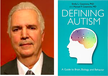
He is the author of several books about autism, including the recent “Defining Autism: A Guide to Brain, Biology, and Behavior,” with Emily L. Casanova, PhD (Jessica Kingsley Publishers, 2019). Dr. Casanova also writes a popular blog corticalchauvinism.com that offers information about autism.
Editor’s note: It Takes Brains to Solve Autism
Because post-mortem brain tissue is needed to study brain structure and development in autism, you might consider registering with Autism BrainNet. This is a research program where families can later make the heroic decision to donate brain tissue in the event of death of an autistic loved one.
References
Andrews DS, Avino TA, Gudbrandsen M, et al. In vivo evidence of reduced integrity of the gray-white matter boundary in autism spectrum disorder. Cerebral Cortex 27(2):877-887, 2017.
Casanova MF, Switala AE, Trippe J, Fitzgerald M. Comparative minicolumnar morphometry of three distinguished scientists. Autism 11(6):557-69, 2007.
Casanova MF. The neuropathology of autism. In Fred Volkmar, Kevin Pelphrey, Rhea Paul, Sally Rogers (eds). Handbook of Autism and Pervasive Developmental Disorders 4th edition, ch. 21. Pp. 497-531, 2014.
Casanova MF, Sokhadze E, Opris I, Wang Y, Li X. Autism spectrum disorders: linking neuropathological findings to treatment with transcranial magnetic stimulation. Acta Paediatrica 104(4):346-55, 2015.
Casanova MF, Casanova EL. The modular organization of the cerebral cortex: evolutionary significance and possible links to neurodevelopmental conditions. Journal of Comparative Neurology doi: 10.1002/cne.24554. [Epub ahead of print] Review.
Wegiel J, Kuchna I, Nowicki K, et al. The neuropathology of autism: defects of neurogenesis and neuronal migration, and dysplastic changes. Acta Neuropathol 11:755-770, 2010


